This 3D human could revolutionize precision medicine and make animal testing obsolete
Parametric Human Project is building the Wikipedia of the human body

Imagine having a map of all the human anatomical parts and their possible variations: a digital collection of all the different types of bones, muscles and connective tissue in men and women from any race. And imagine that a high-speed computer could download personal data into this 3D simulated human and run tests for doctors to determine exactly how a patient will respond to a particular drug or treatment without first testing it on animals.
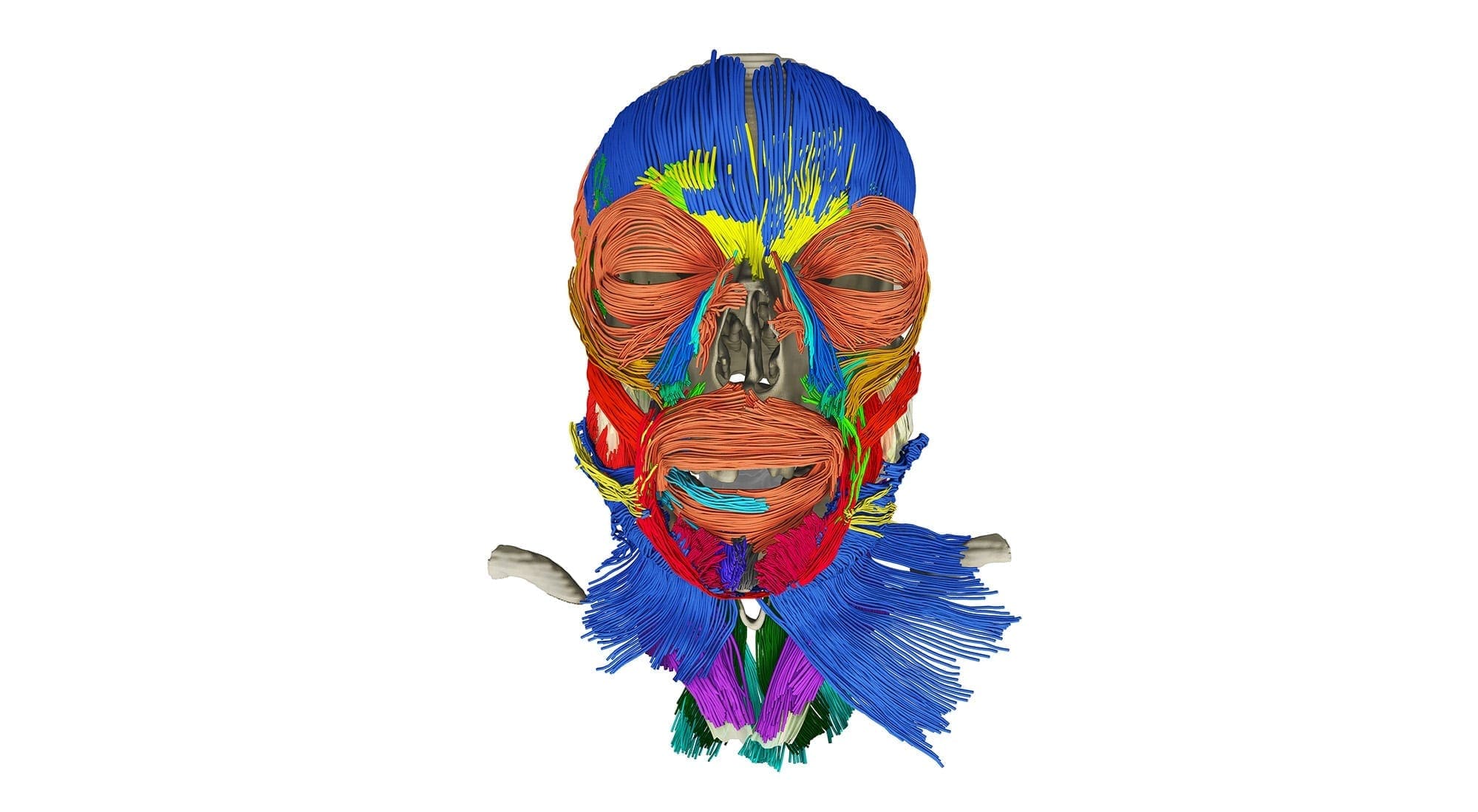
The promise of precise, customized medicine is still years away, but a cross-disciplinary team, involving 30 institutions around the world, is building a complete morphological and physiological virtual human dubbed the Parametric Human.
Accurate diagnosis and treatment rely on solid information, yet “we really don’t know the human body,” says Jeremy Mogk, Autodesk Research’s principal scientist on the project.

“Even when you look at anatomy textbooks, what they’re really showing is a relatively superficial view,” he adds. Mogk believes it’s time to go beyond the superficial — by graphically depicting, in minute detail, every part of the body, which is why he has spearheaded the project through Autodesk, one of the world’s largest software design firms.
But despite backing by both corporate and academic partners, intense effort is required. Mr. Mogk points to work under way at the University of Toronto, where Prof. Anne Agur of the department of surgery is digitizing muscle fibres, section by section, throughout the body with a miniature robotic arm.

“Through the digitization process that she pioneered, she’s discovering that we had no idea just how complex some of these structures are,” says Mr. Mogk.
The project’s overarching goal, he adds, is to create a central repository and platform where researchers can share their data — a Wikipedia, of sorts, for all things related to the human body. But the potential applications are no less exciting: the 3D model can be used to develop targeted therapies, to map complex surgeries and to create simulation-based training for healthcare professionals.
“By creating this model, we are building something that can be used as a reference or map of the human body, complete with all the possible variations so that, for example, surgeons know what to expect when they’re going into a particular bone or joint,” says Mr. Mogk.
![]()
 Marjo Johne